
By D. Brant. Marymount College.
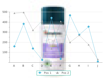
Many of them are potent anticonvulsants discount proscar 5 mg overnight delivery, especially when tested against PTZ and retard the development of kindling buy proscar 5mg online. Unfortunately their clinical value is limited by the development of tolerance cheap proscar 5 mg mastercard. Barbiturates also potentiate the action of GABA but as they can do this by directly increasing the duration of opening of the chloride ion channel proscar 5mg online, independently of the GABA or benzadiazepine receptor sites, they cannot strictly be considered to augment GABA. Glutamate NMDA receptor antagonists such as AP5 and AP7 were first shown to be anticon- vulsant following introcerebroventricular injection into DBA/2 mice susceptable to audiogenic seizures. In addition, they offer protection to PTZ, reduce the after- discharge in amygdala kindled rats and can actually retard the development of kindling. Although AP7 has some effect in photosensitive baboons, systemically active com- pounds have proved difficult to synthesise. Recently felbamate, an antagonist at the glycine-sensitive site on the NMDA receptor, has shown systemic anticonvulsant activity and clinical efficacy. Inhibition of glutamate release was thought to be the mode of action of lamotrigine. It reduces MES and kindling and also glutamate (and to a lesser extent GABA) release induced in brain slices by veratridine, which opens sodium channels. But it now seems likely that the actual block of sodium channels is its primary action (see later). The epileptic discharges induced in hippocampal slices by tetanic stimulation has been shown to be accompanied by reduced GABA-mediated IPSPs (Stelzer, Slater and Bruggencate 1987). Since AP7 not only reduced the discharges but also restored the response to GABA some linkage between NMDA and GABAA receptors seems probable. In fact the interaction between glutamate and GABA probably means that both of them and possibly their different receptors may need to be manipulated appro- priately to control convulsive activity. This has been shown in fact experimentally when bicuculline was infused intravenously for short periods in the rat to give a burst of epileptic-like spiking in the EEG. Superfusion of the cortex using the cup technique with the glutamate AMPA antagonist CNQX or the GABAB agonist baclofen reduced the actual number (initiation) of spikes but not their amplitude, while NMDA antagonists (AP7) and the GABAA agonist muscimal reduced the size (development and spread of excitation) and not the number of spikes (Zia-Gharib and Webster 1991). Clearly more than one aspect of amino acid function may need to be controlled. Other NTs have been implicated in the aetiology of epilepsy but direct evidence is lacking. Many studies have also shown that cortical ACh release increases in proportion to EEG activity during the administration of a wide range of convulsants. Nevertheless while cholinergic-induced seizures can be suppressed by antimuscarinic drugs they have no effect against any epilepsy in humans and ACh release presumably reflects rather than directly causes cortical activity. MONOAMINES The widespread and diverging nature of ascending monoamine pathways to the cortex suggest that NA and 5-HTare more likely to have a secondary modifying rather than a primary effect on the initiation of epileptic activity. In reality this is the case and their secondary role is even a minor one. Generally a reduction in monoamine function facilitates experimentally induced seizures (see Meldrum 1989) while increasing it reduces seizure susceptibility. The variability of the procedures used and results obtained do not justify more detailed analysis here. Some mention should perhaps be made of dopamine, considering its role in the control of motor function. It is perhaps not surprising that DA agonists like apo- morphine block the myoclonus induced in photosensitive baboons and audiogenic seizures in DBA/2 mice while neuroleptics (DA antagonists) may have a weak procon- vulsant effect in humans. Also in rats with absence seizures dopa, apomorphine and D1 agonists reduce facial clonus and spike and wave discharges, while the D1 antagonist SCH 23390 increases them. Nevertheless, there is no evidence of a significant role for DA (or NA and 5-HT) in human epilepsies. ADENOSINE A number of studies have shown that adenosine inhibits neuronal firing both in vitro and in vivo and is itself released during intense neuronal activity. It can protect against PTZ seizures in rodents while the antagonist theophylline is proconvulsant. APPROACHES TO THE CONTROL OF EPILEPTIC ACTIVITY Irrespective of the cause of epilepsy, the spread of seizure activity will be attenuated by either decreasing the excitation or increasing the inhibition of neurons. This may be achieved in a number of ways, either directly by (a) blocking excitatory voltage-gated Na (or possibly Ca2) channels (1) (b) increasing the opening of inhibitory Cl7 channels (2) or indirectly by (c) reducing the release of the excitatory NT, glutamate (3) or its action at NMDA receptors (4) 342 NEUROTRANSMITTERS, DRUGS AND BRAIN FUNCTION (d) increasing the availability (and release) of the inhibitory NT, GABA by blocking its reuptake (5) or metabolism (6) or activating the GABA receptor either directly (7) or through the benzodiazepine receptor (8).
Injury to the gracile fasciculus on the right would result is other types of motor deficits buy proscar 5mg without prescription. The level of the cord damage is caudal to the cuneate fasciculi and the anterolateral system con- 38 safe proscar 5mg. Answer A: The exiting fibers of the abducens nerve (on the left) veys pain and thermal sensations cheap 5mg proscar. Answer A: The loss of pain and thermal sensations on the right Diplopia may result from lesions of the oculomotor and trochlear side of the body correlates with a lesion involving the anterolateral nerves proscar 5 mg on-line, but these structures are not in the domain of the parame- system on the left side of the spinal cord. A lesion of the optic nerve results in blind- terolateral system would result in a left-sided deficit. The gracile ness in that eye and damage to the facial root does not affect eye and cuneate fasciculi convey discriminative touch, vibratory sen- movement but may cause a loss of view of the external world if the sation, and proprioception. The posterior spinocerebellar tract palpebral fissure is closed due to facial muscle weakness. Answer E: The substantia nigra contains a large population of dial lemniscus is located within the territory served by paramedian melanin-containing cells, is located in the midbrain just internal to branches of the basilar artery. Penetrating branches of the anterior the crus cerebri, and the loss of these cells gives rise to the motor spinal artery serve the hypoglossal nucleus. The neurotransmitter generally in the territories of short or long circumferential associated with these cells is dopamine. Answer D: Weakness of the extremities accompanied by paralysis cleus is in the midbrain, but its reddish tone is related to a rich vas- of the lateral rectus muscle (innervated by the abducens nerve) on the cular supply, not to cells containing a pigment. Answer E: The solitary nucleus is located immediately inferior middle alternating hemiplegia. Inferior alternating hemiplegia speci- (ventral) to the medial and spinal vestibular nuclei and is the only fies involvement of the hypoglossal root and the pyramid, and supe- nucleus in the choices to receive a general visceral afferent (GVA) rior alternating hemiplegia indicates damage to the oculomotor root and special visceral afferent (SVA-taste) input. Alternating (or alternate) hemianesthesia and tory nucleus and the nucleus ambiguus are visceromotor (general hemihypesthesia are sensory losses. Answer B: The prominent elevation formed on the caudal and (general somatic afferent [GSA] and special somatic afferent [SSA], medial wall of the third ventricle, at the general level of the pos- respectively). Answer E: The superior salivatory nucleus lies adjacent to the ignating the separation between the third ventricle (rostral to this exiting fibers of the facial nerve in a position just lateral to the ab- point on the midline) and the quadrigeminal cistern (caudal to this ducens nucleus in caudal levels of the pons. The pulvinar is lateral to the quadrigeminal cistern, the ons originating from these cells distribute on peripheral branches lamina terminalis forms the rostral wall of the third ventricle, and of the facial nerve. The dorsal motor and inferior salivatory nuclei the massa intermedia bridges the space of the third ventricle. The Edinger-Westphal nucleus is related pears as a shadow in T2-weighted MRI bridging the third ventri- to the oculomotor nucleus and the intermediolateral cell column cle. The superior colliculus is a mesencephalic structure found in is located primarily in thoracic levels of the spinal cord. Answer C: The oculomotor nucleus (containing general somatic nerve, portions of the corticospinal fibers in the crus cerebri, and efferent [GSE] cell bodies), along with the Edinger-Westphal (con- a number of other medially located structures are found in the ter- taining general visceral efferent [GVE] cell bodies) nucleus, is ritory of the penetrating branches of the basilar bifurcation. The found in the most anterior and medial portion of the periaqueduc- paramedian branches of the basilar artery and the corticospinal tal grey at the superior colliculus level. The trochlear nucleus is fibers in the pyramid serve the abducens nerve by branches of the found at a comparable position, but at the cross-sectional level of anterior spinal artery. The mesencephalic nucleus is found in the lemniscus are mainly, if not entirely, in the region of the midbrain lateral area of the periaqueductal grey, and the trigeminal and ab- served by branches of the quadrigeminal and posterior medial ducens nuclei are located in the pons. Answer D: The vocalis muscle (this muscle is actually the me- acterized by a loss of most eye movement (damage to oculomotor dial portion of the thyroarytenoid muscle) is innervated, via the nerve fibers) on the ipsilateral side and weakness of the upper and vagus nerve, by motor neurons located in the nucleus ambiguus. The abducens nerve is the cranial nerve in- spinal trigeminal nucleus relays sensory input from the face. The volved in a middle alternating hemiplegia and the hypoglossal is that hypoglossal nucleus is motor to the tongue and the facial nucleus nerve involved in an inferior alternating hemiplegia. Answer A: Fibers comprising the anterolateral system convey pain and thermal sensations from the body, excluding the face. Answer E: Motor neurons in the nucleus ambiguus innervate, These fibers are located in lateral portions of the medulla adjacent primarily through the vagus nerve, the muscles of the throat that to the spinal trigeminal tract; this latter tract relays pain and ther- move a bolus of food from the oral cavity to the esophagus. The gracile and cuneate fasciculi con- tongue, via the hypoglossal nucleus and nerve, may move food vey proprioception, discriminative touch, and vibratory sense in around in the mouth and toward the back of the oral cavity, but the spinal cord and the medial lemniscus conveys this same infor- the actual act of swallowing is through the action of pharyngeal and mation from the medulla to the dorsal thalamus.
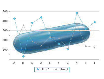
GnRH binds to high- affinity receptors on the gonadotrophs and stimulates the FSH/LH PRL secretion of LH and FSH through a phosphoinositide-pro- tein kinase C-mediated pathway (see Chapter 1) 5 mg proscar fast delivery. Inhibin cheap proscar 5mg without a prescription, activin cheap 5 mg proscar with visa, A graph of LH release throughout the female life span is Ovary follistatin shown in Figure 38 purchase 5mg proscar mastercard. During the neonatal period, LH is re- leased at low and steady rates without pulsatility; this pe- riod coincides with lack of development of mature ovarian Estradiol, follicles and very low to no ovarian estradiol secretion. Pul- progesterone, androgen satile release begins with the onset of puberty and for sev- eral years is expressed only during sleep; this period coin- cides with increased but asynchronous follicular Reproductive Secondary sex development and with increased secretion of ovarian estra- tract characteristics diol. Upon the establishment of regular functional men- Regulation of the reproductive tract in the strual cycles associated with regular ovulation, LH pulsatil- FIGURE 38. The main reproductive hormones are ity prevails throughout the 24-hour period, changing in a shown in boxes. Positive and negative regulations are depicted by monthly cyclic manner. The ovaries are in the pelvic portion of the abdominal cav- ity on both sides of the uterus and are anchored by ligaments THE FEMALE REPRODUCTIVE ORGANS (Fig. An adult ovary weighs 8 to 12 g and consists of an outer cortex and an inner medulla, without a sharp demarca- The female reproductive tract has two major components: tion. The cortex is surrounded by a fibrous tissue, the tunica the ovaries, which produce the mature ovum and secrete albuginea, covered by a single layer of surface epithelium progestins, androgens, and estrogens; and the ductal sys- continuous with the mesothelium covering the other organs tem, which transports ovum, is the place of the union of the in the abdominal cavity. The cortex contains oocytes en- sperm and egg, and maintains the developing conceptus closed in follicles of various sizes, corpora lutea, corpora al- until delivery. The morphology and function of these struc- bicantia, and stromal cells. The medulla contains connective Isthmus Fundus Ampulla Corpus Broad ligament Uterus Oviduct Myometrium Fimbria Endometrium Ovary Infundibulum Primordial follicle Cervix Primary follicle Vagina Ovarian ligament Atretic follicle Ovarian vessels Early antrum formation Corpus albicans Mature corpus luteum Ovary Graafian follicle Early corpus luteum Stroma Germinal epithelium Ovulation FIGURE 38. Blood vessels, lymphatics, and nerves cation (keratinization) of the vaginal epithelium, whereas enter the medulla of the ovary through the hilus. The Estradiol also activates vaginal glands that produce lubri- oviducts are the site of fertilization and provide an envi- cating fluid during coitus. The oviducts are 10 to 15 cm long and composed of sequential regions called the infundibulum, ampulla, and isthmus. FOLLICULOGENESIS, STEROIDOGENESIS, The infundibulum is adjacent to the ovary and opens to the ATRESIA, AND MEIOSIS peritoneal cavity. It is trumpet-shaped with finger-like pro- Most follicles in the ovary will undergo atresia. However, jections called fimbria along its outer border that grasp the some will develop into mature follicles, produce steroids, ovum at the time of follicular rupture. As follicles mature, oocytes will also mature by ered with densely ciliated projections, which facilitate entering meiosis, which produces the proper number of ovum uptake and movement through this region. The isthmus is located at the uterotubal junction and has a narrow lumen surrounded The Primordial Follicle Contains an by smooth muscle. It has sphincter-like properties and can Oocyte Arrested in Meiosis serve as a barrier to the passage of germ cells. The oviducts transport the germ cells in two directions: sperm ascend to- Female germ cells develop in the embryonic yolk sac and ward the ampulla and the zygote descends toward the migrate to the genital ridge where they participate in the uterus. This requires coordination between smooth muscle development of the ovary (Table 38. Without germ contraction, ciliary movement, and fluid secretion, all of cells, the ovary does not develop. Oogonia undergo mi- The uterus is situated between the urinary bladder and tosis only during the prenatal period. On each upper side, an oviduct opens into the uter- contain a finite number of oocytes, estimated to be about 1 ine lumen, and on the lower side, the uterus connects to the million. The puberty, only 200,000 oocytes remain; by age 30, only outer part is the myometrium, composed of multiple layers 26,000 remain; and by the time of menopause, the ovaries of smooth muscle.